2. 东北师范大学生命科学学院分子表观遗传学教育部重点实验室, 长春 130024
2. School of Life Sciences, Northeast Normal University, Changchun 130024, China
Grimmia ulaandamana J. Muñoz, C. Feng, X.L. Bai & J. Kou was recently described from several localities in China[1]. The species is mainly characterized by having 2-4(5)-stratose, v-shaped lamina, semiterete to terete costa consisting of almost homogeneous cells and with four guide cells at mid-leaf, and a long plication in lower part on one or both sides of the leaf. Even though G. ulaandamana was described based on several specimens in different herbaria, sporophytes are very rarely produced, only one sporophyte attached to the plant was available, and it was old and in such a poor condition. Another complete capsule was found among the debris of one specimen, but without connection to the gametophytes and without seta[1]. Obviously, it is not adequate for description.
During the field trip in Saihanwula National Nature Reserve and adjacent areas in 2013, the second author made a large collection of bryophytes and more than 500 specimens were collected. In the process of identification several fertile populations of G. ulaandamana were discovered. The characteristics of the mature sporophytes of G. ulaandamana, as well as both female plants and male plants are described here.
1 Material and methodsThe specimen was studied with the anatomical and morphological methods applied for the mosses[2]. Specimen collected is deposited at BAU. Microscopic examinations and measurements were taken with a ZEISS Primo Star light microscope, while microphotographs were obtained with a Canon EOS 70D camera mounted on this microscope. Specimens were examined in 2% KOH.
2 ResultsGrimmia ulaandamana J. Muñoz, C. Feng, X.L. Bai & J. Kou. (Figs. 1–3)

|
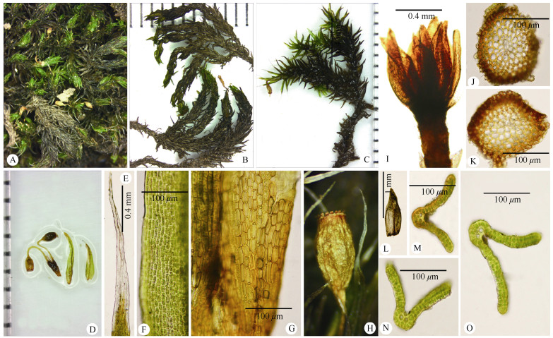
Fig. 1 Gametophyte and sporophyte of Grimmia ulaandamana. A: Plants; B: Plants when dry, female above, male below; C: Plants when moist; D: Sporophytes (left), innermost perichaetial leaf (middle) and cauline leaf (right); E: Hair-points of innermost perichaetial leaf; F: Mid-leaf cells of innermost perichaetial leaf; G: Basal part of innermost perichaetial leaf; H: Capsule when dry; I: Perigonium. J, K: Cross-sections of the same stem (male plant). L: Innermost perigonial leaves. M-O: Cross-sections of leaves. (from Li Feng 2013018, NMAC). |

|
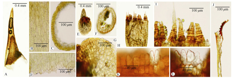
Fig. 2 Sporophyte of Grimmia ulaandamana. A: Calyptra; B: Middle part of calyptra; C: Cross-sections of calyptra (middle part); D: Basal part of calyptra; E: Operculum; F: Cross-section of operculum; G: Basal cells of operculum; H-J: Peristome teeth; K, L: Annulus. (from Li Feng 2013018, NMAC). |

|
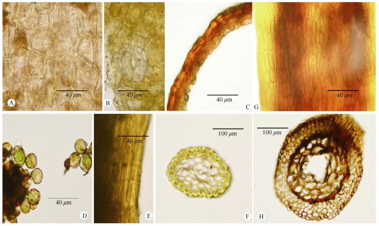
Fig. 3 Sporophyte of Grimmia ulaandamana. A: Exothecial cells of capsule (middle portion); B: Stomata; C: Cross-sections of urn (middle part); D: Spores; E: Exothecial cells of seta; F: Cross-section of seta; G: Exothecial cells of vaginula; H: Cross-section of vaginula. (from Li Feng 2013018, NMAC). |
Type: CHINA: Inner Mongolia, Ulaan dam-Shipeng Ditch National Nature Reserve, Wanghuolou Mountain, 44°26ʹ519ʺ N, 118°45ʹ955ʺ E, on rocks, mixed with Macromitrium japonicum, 1 967 m, 12 July 2011, Chao Feng 2011527-2. Holotype: HIMC (seen); isotype: MA-Musci 40053.
Plants dioicous. Female plants in dark green, loose, not hoary, readily disintegrating tufts; stem central strand consistently present; leaves spreading when moist; innermost perichaetial leaves enlarged, about 1.5 times longer than cauline leaves, 3.1–3.5 mm long, oblong-lanceolate to long-lanceolate with a hairpoint, longitudinally plicate in lower half, sheathing, with a percurrent costa, with areolation of rectangular cells with moderately thickened walls, straight. Male plants similar to but differs from female plants in less robust plants and stem central strand weak or absent, irregular development in the same stem; perigonia gemmiform; innermost perigonial leaves smaller than cauline leaves, broadly ovate, concave, broadly acute, muticous, with a thin-walled areolation and a percurrent costa. Sterile plants similar to female plants. Setae usually 2 per perichaetium, 1.3–1.8 mm long, yellowish to brown when mature, arcuate, cells rectangular, thick-walled, in cross-section with 1(–2) layers of thick-walled, strongly bulging cortical cells and a group of thin-walled medullar cells surrounding a differentiated central strand. Vaginula 0.88–1 mm long, made up of nearly rectangular cells with curved, thin, smooth walls, not tortion, in cross-section with 4–5 layers of thin to slightly thick-walled, ovate, concentric cells. Capsules exserted, pendent, obloid, 0.78– 1.18 mm×0.33–0.45 mm, striate, dull, dark yellow, in cross-section exterior wall slightly bulging, thickwalled. Annulus conspicuous, deciduous, made up of 2 rows of pellucid, thick-walled cells, rectangular in upper row, quadrate to short-rectangular in lower row (elongata-type). Operculum convex, with a short, straight beak, base uneven, formed by nearly isodiametric cells. Exothecial cells irregular in shape, from quadrate to short rectangular or hexagonal, thick-walled, 20–32.5 μm×(7.5–)12.5–20 μm. Stomata present in the base of the capsule. Peristome teeth lanceolate, 0.15– 0.2 mm long, perforated in upper half, at dehiscence joined at base, trabeculae broader in lower part, dorsal lower plates smooth, upper half of dorsal side and ventral side densely covered with coarse papillae, in longitudinal section, teeth inserted below orifice, 1 layer of small cells between exothecium and teeth present, trabeculae close together, in lower third markedly protruding, in upper part scarcely protruding. Spores 9–17 μm diameter, spherical, finely granulose. Calyptra cucullate, smooth, in cross-section composed of 3–4 layers of thick-walled cells in the middle part.
Specimen examined: CHINA: Inner Mongolia: Saihanwula National Nature Reserve, Hexinqu: 44° 25.142′ N, 118°64.793′ E, on rock, ca 1 523 m, 10 July 2013, Li Feng 2013018, Li Feng 2013056, Li Feng 2013060, Li Feng 2013078 (NMAC).
Distribution and habitat: Grimmia ulaandamana is only known from several districts in China: Heilongjiang, Inner Mongolia, Hebei, Shaanxi and Yunnan, currently considered endemic to China. These plants were found on rocks, as ever, mixed with Macromitrium japonicum Dozy & Molk. Female plants bearing mature sporophytes and male plants grew close together.
3 Discussion 3.1 VariationThe presence or absence of stem central strand is usually treated as a diagnostical character in the mosses taxonomy, although there are exceptions in limited genera or species[2]. In the protologue, the stem central strand was described as present in G. ulaandamana[1]. However, the strand development is correlated with sexual maturity in the light of the present observation. In addition to G. ulaandamana, we also found that stems of G. pilifera P. Beauv. and G. elatior in this area are characterized by stem central strand irregular developement in the same stem. This case was also noted by Maier and the same situation may occurred in other species of Grimmia, such as G. atrata Miel. ex Hornsch[3]. Although the presence or absence of the stem central strand is usually variable in some species of Grimmia, this should not be neglected because it is rather constant and of taxonomic importance in other species[4]. For example, the stem central strand of G. pilifera is usually absent, and is diagnostically useful to recognize this species in most situations, especially when the leaf shape and laminal stratification of G. pilifera are usually highly variable.
The upper laminal stratification of G. ulaandamana is usually variable from bistratose with unistratose patches to 4(5)-stratose. In Grimmia, some other species such as G. elatior, G. longirostris Hook., G. ovalis (Hedw.) Lindb., G. pilifera, G. unicolor Hook. can possess the capacity to have a variable laminal stratification. The multistratose condition may occur when these plants grow in proper amount of moisture environment[5]. However, like stem central strand, the laminal stratification is rather constant and of taxonomic importance in other species[4].
Four guide cells at mid-leaf and two to four at leaf-insertion were described in G. ulaandamana[1]. In the present study, the costa at leaf-insertion is usually narrow with two or four guide cells, but may be occasionally wider, consisting of two or four guide cells and a double layer of companion cells (smaller than guide cells and with more thickened walls) in the ventral side of costa. This case may occur in other species of Grimmia such as G. pilifera and G. elatior from our observation. As a consequence, examination of the costal guide cells at leaf-insertion should be more careful to distinguish them from other ventral cells of costa. In addition, number of costal guide cells at leaf-insertion was advocated as critical by Maier[3] and we agree that accurately counting the number of such cells is sometimes difficult[6], partly owing to the two outer guide cells of costa at leaf-insertion usually contiguous with laminal basal cells or unsharp boundary of costa at this position. In addition, the number of guide cells within certain species is usually variable from six to eight or from four to six. Nevertheless, its number may still be an important clue when differentiating similar species of Grimmia. Recently, Delgadillo-Moya[7] retained the "ventral cell" in Grimmia instead of "guide cells" to distinguish them from the guide cells of other moss group. Indeed, the words "guide cells" in other mosses groups such as Echinodiaceae, Dicranaceae or Pottiaceae refer to the thin-walled, large cells usually present at the middle position of costa or just below the differentiated ventral epidermis cells. However, the words "ventral cell" seem to be unsuitable for genus Grimmia to replace "guide cells" because of the occasional presence of companion cells on the ventral surface of costa which are not large and thinwalled.
3.2 Phylogenetic implicationsThe discovery of mature sporophytes of G. ulaandamana indicates that it is not the male plants of G. elatior, and the comparison with the latter species on gametophyte was in Feng et al.[1]. The additional comparison mainly on sporophyte was summaried in Table 1. While the presence or absence of laminal papillose of G. elatior was differently described from different regions[3–4, 8–10], both of these plants with or without papillose of G. elatior were found in the same area. Grimmia ulaandamana also grew in the same area where G. elatior was abundant. The large size, bulging upper laminal cells, narrowly furrowed and irregularly angled costa in cross section make G. elatior easy to be distinguished.
With the discovery of mature sporophytes and according to Cao et al.[8], Muñoz[11] and Hastings et al.[4], the presence of stem central strand, sharply keeled and lanceolate to ovate-lanceolate leaves, costa projecting on abaxial side, leaf margin recurved on one or two sides, long, arcuate to cygneous seta when moist, symmetric, pendulous and ribbed capsule, stomata present at base of capsule and cucullate calyptra suggest the placement of G. ulaandamana in the Grimmia subgen. Rhabdogrimmia Limpricht. According to Ochyra et al.[12], Grimmia was split into seven genera. Among them Dryptodon Brid. included the species formerly treated as Grimmia subg. Rhabdogrimmia, and G. ulaandamana therefore may belong to this genus at this point.
In the subgen. Rhabdogrimmia, Grimmia elatior Bruch ex Balsamo-Crivelli & De Notaris is most similar to G. ulaandamana. Both species share similar laminal areolation, 2-4(5)-stratose lamina and the costa protruding dorsally from the lamina, undifferentiated cells of the costa in cross-section, arcuate seta, exserted and striate capsule. Although G. ulaandamana could be distinguished from the female plants of G. elatior in several aspects stated in Feng et al.[1], the differences between G. ulaandamana and male plants of G. elatior are very minimal because the latter has semi-terete to nearly terete costa, usually smooth upper laminal cells and rectangular juxtacostal cells with straight walls. The only noteworthy difference between them is present in their different leaf posture when moist. The comparison between G. ulaandamana and G. elatior on most gametophyte characters was in Feng et al.[1], and the differences on additional gametophyte characters and sporophyte characters were summaried. The leaf posture of G. ulaandamana when moist is patent to spreading, but G. elatior is erect to erecto-patent. About the stem central strand of female plants, G. ulaandamana is consistently present, and G. elatior is present above and absence below. The innermost perichaetial leaves of G. ulaandamana is enlarged, but G. elatior is not enlarged. The exothecial cells of G. ulaandamana is thickened walls, but G. elatior is thin walls. The calyptra of G. ulaandamana is cucullate, not lobed and eroded at base, but G. elatior is mitrate and lobed at base. The peristome teeth of G. ulaandamana is split in upper half, separated above the insertion, trabeculae large in lower, teeth inserted below orifice, 1 layers of small cells between exothecium and teeth, trabeculae in lower third strongly protruding, but G. elatior is deeply split, separated down to the insertion, trabeculae small in lower, teeth inserted at orifice, 1 or 2 layers of large cells between exothecium and teeth, trabeculae in lower third scarcely protruding. The discovery of mature sporophytes indicates that G. ulaandamana is not the male plants of G. elatior, and provides additional evidences to accept this newly described species as distinct.
Another species in subgen. Rhabdogrimmia with which G. ulaandamana could be confused is G. funalis (Schwägr.) Bruch & Schimp. The former occasionally has 2-4 stratose laminal cells and costa weak proxymally, projecting on abaxial side. Nevertheless, G. funalis differs from G. ulaandamana by its leaves patent when moist, upper and median laminal cells with strongly thickened and moderately sinuose walls, peristome teeth inserted near orifice and mitrate calyptra.
Acknowledgments We are grateful to Dr Jesús Muñoz, Real Jardín Botánico, for providing important literature. We thank Li Feng to collect the specimens.
| [1] |
FENG C, MUÑOZ J, KOU J, et al. Grimmia ulaandamana (Grimmiaceae), a new moss species from China[J]. Ann Bot Fenn, 2013, 50(4): 233-238. DOI:10.5735/086.050.0405 |
| [2] |
ZANDER R H. Genera of the Pottiaceae: mosses of the harsh environments[J]. Bull Buffalo Soc Nat Sci, 1993, 32: 1-378. |
| [3] |
MAIER E. The genus Grimmia Hedw. (Grimmiaceae, Bryophyta): A morphological-anatomical study[J]. Boissiera, 2010, 63: 1-377. |
| [4] |
HASTINGS R I, GREVEN H C. Grimmia Hedw [M]// Flora of North America north of Mexico, Vol. 27. New York: Oxford University Press, 2007: 225–258.
|
| [5] |
DEGUCHI H. A revision of the genera Grimmia, Schistidium and Coscinodon (Musci) of Japan[J]. J Sci Hirosh Univ Ser B Div Bot, 1978, 16(2): 121-256. |
| [6] |
MILLER N G, HASTINGS R I. Taxonomy and distribution of Grimmia (Bryophyta) in mountain regions of the northeastern United States[J]. Bryologist, 2013, 116(1): 28-33. DOI:10.1639/0007-2745-116.1.028 |
| [7] |
DELGADILLO-MOYA C. Grimmia (Grimmiaceae, Bryophyta) in the Neotropics [M]. México: Univ Nac Autón Méx, 2015: 8–81.
|
| [8] |
CAO T, HE S, VITT D H. Grimmiaceae [M]// GAO C, CROSBY M R, HE S. Moss Flora of China, Vol. 3. Beijing: Science Press & St. Louis: Missouri Botanical Garden Press, 2003: 3–76.
|
| [9] |
IGNATOVA E A, MUÑOZ J. The genus Grimmia (Grimmiaceae, Musci) in Russia[J]. Arctoa, 2005, 13(1): 101-182. DOI:10.15298/arctoa.13.13 |
| [10] |
MAIER E. The genus Grimmia (Musci, Grimmiaceae) in the Himalaya[J]. Candollea, 2002, 57: 143-238. |
| [11] |
MUñOZ J. A taxonomic revision of Grimmia subgenus Orthogrimmia (Musci, Grimmiaceae)[J]. Ann Mo Bot Gard, 1998, 85(3): 367-403. DOI:10.2307/2992039 |
| [12] |
OCHYRA R, ŻARNOWIEC J, BEDNAREK-OCHYRA H. Census Catalogue of Polish Mosses[M]. Kraków: Pol Acad Sci, Inst Bot, 2003: 120-125.
|
 2023, Vol. 31


